"The Anatomy Lesson of Dr. Nicolaes Tulp," detail, by Rembrandt, 1632. Mauritshuis, Den Haag. Image is in the public domain.
1. INTRODUCTIONCh. 1 Introduction: structure & function of the body
Ch. 2 Chemistry of life Ch. 3 Cells Ch. 4 Tissues |
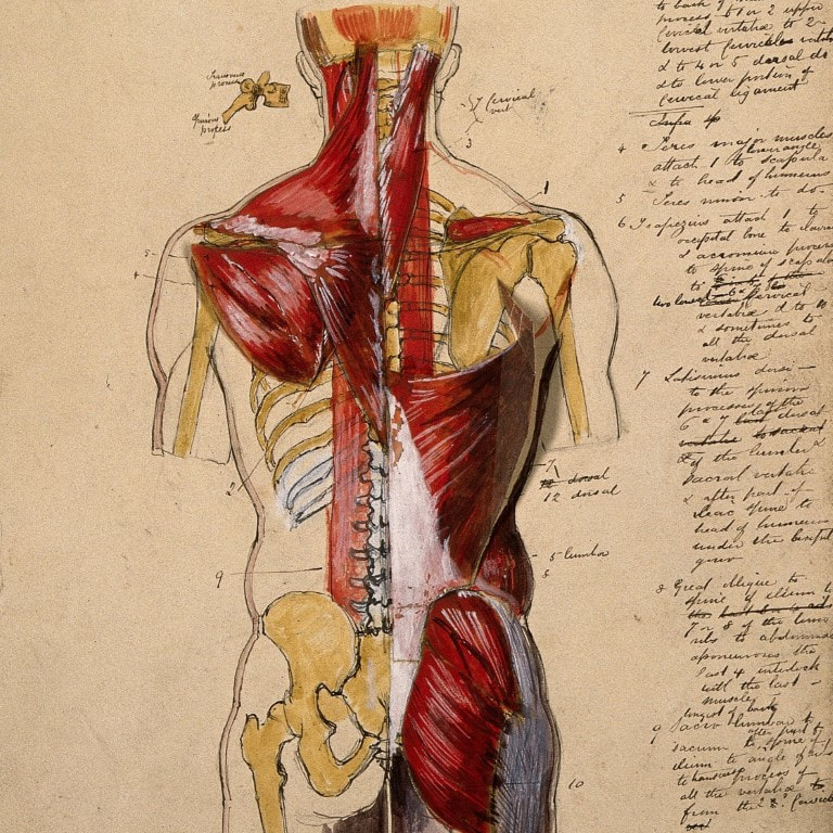
"Dissection of the trunk," ink and watercolour, 18-- . Wellcome Collection. Image has been cropped and is used under a CC BY 4.0 license.
|
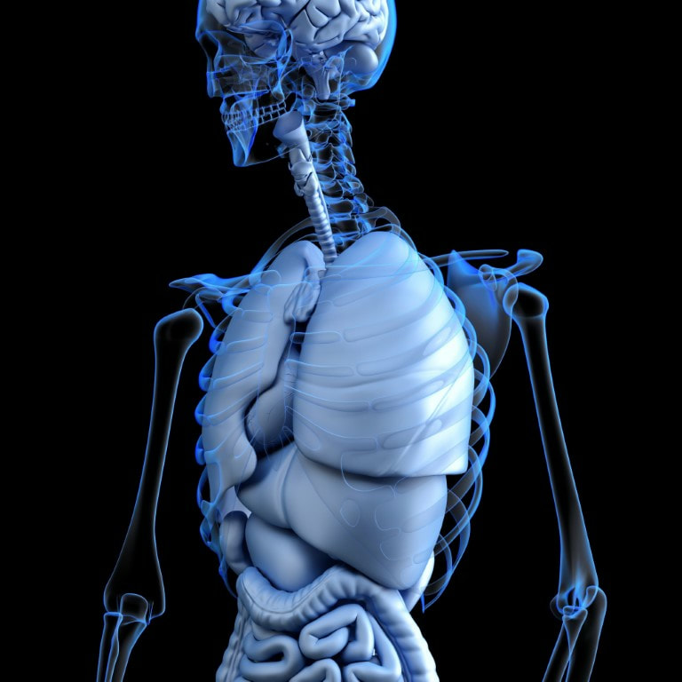
2. DISEASE AND THE BODYCh. 5 Organ systems
Ch. 6 Mechanisms of disease |
"Anatomy" (situs inversus). Image by www_slon_pics has been cropped and is used under a Pixabay license.
|
3. THE INTEGUMENTCh. 7 The Integumentary System & Body Membranes
|
Skin cancer cells - squamous cell carcinoma (NIH Image Gallery). Image by Markus Schober and Elaine Fuchs, The Rockefeller University, used under a CC BY-NC 2.0 license.
|
4. THE MUSCULOSKELETALSYSTEM |
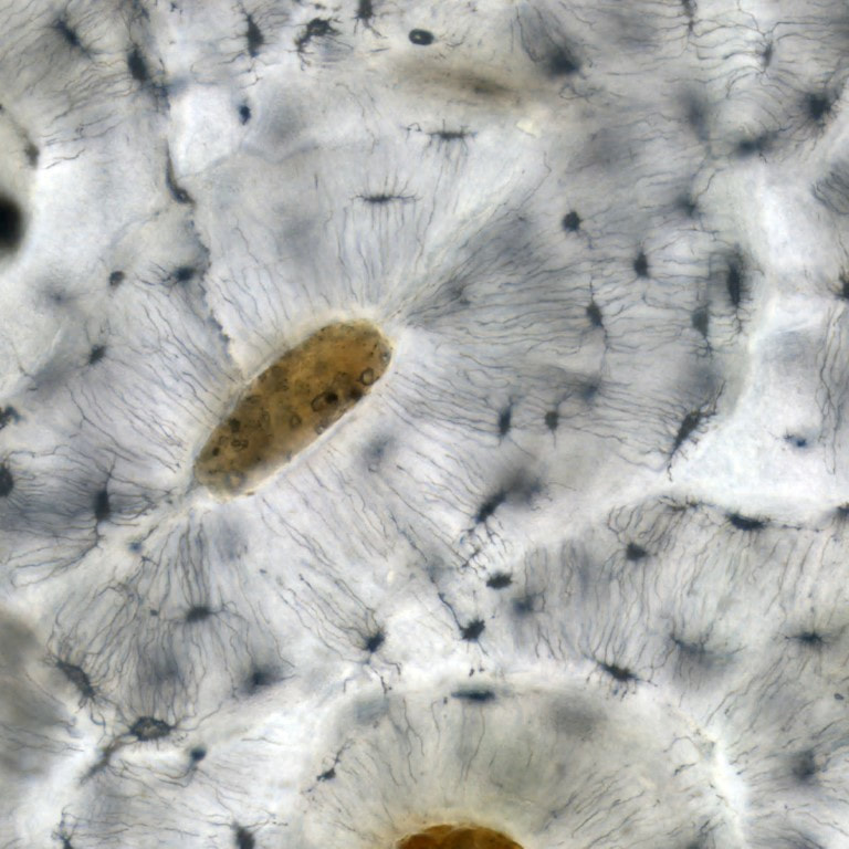
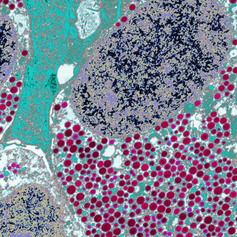
Light micrograph of osteocytes (long black cells), their canaliculi, and haversian canals. Wellcome Collection. Image by Kevin Mackenzie, University of Aberdeen, has been cropped and is used under a CC BY 4.0 license.
|
5. THE NERVOUS SYSTEMAND THE SENSES |
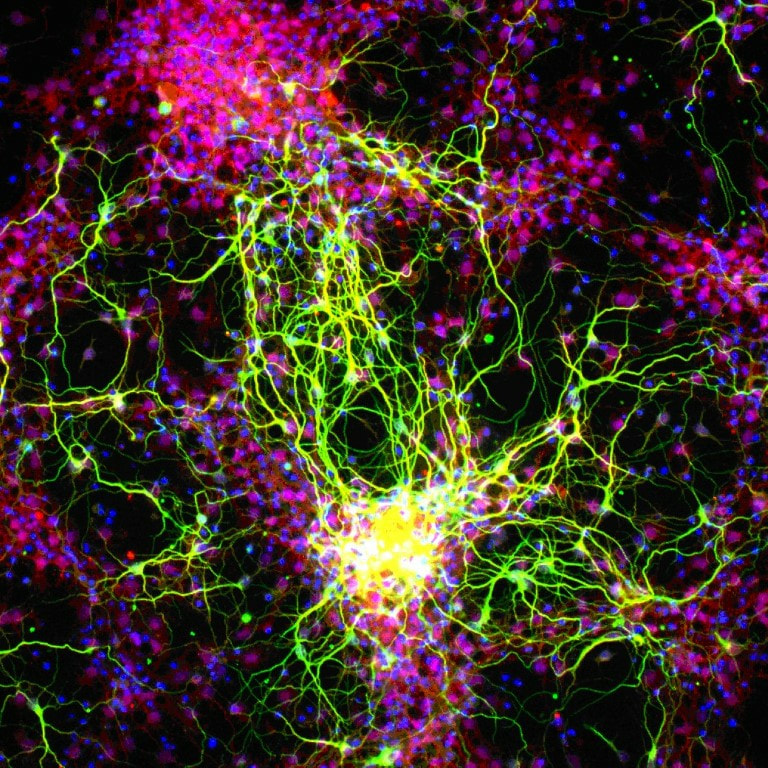
Retinal ganglion cells. Image courtesy of the National Eye Institute, NIH (Courtesy of Thomas V. Johnson, Naoki Nakaya, and Stanislav Tomarev of the NEI Laboratory of Retinal Cell and Molecular Biology, Molecular Mechanisms of Glaucoma Section). Image has been cropped and is used under a CC BY-NC 2.0 license.
|
6. THE ENDOCRINE SYSTEMCh. 12 The Endocrine System
|
Hormone-producing cell from the anterior lobe of the pituitary gland. The cell is full of growth hormone granules. University of Edinburgh. Image has been cropped and is used under a CC BY-NC 4.0 license.
|
7. THE CARDIOVASCULARSYSTEMCh. 13 Blood
Ch. 14 The Heart Ch. 15 The Circulation of the Heart KA - Fetal circulation before birth & after birth |

"Colour-enhanced image of a blood clot. There are many red blood cells and a single white blood cell held together in a meshwork of fibrin. The red blood cells are crenated - the spiky appearence that occurs when they become dehydrated." Francis Crick Institute. Image by Anne Weston has been cropped and is used under a CC BY-NC 4.0 license.
|
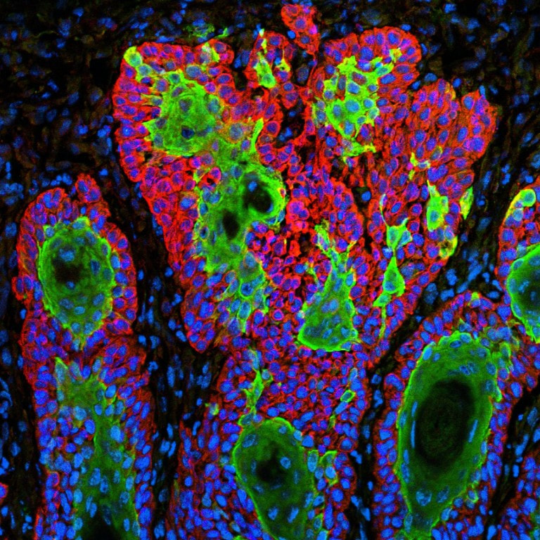
8. THE LYMPHATIC SYSTEMAND IMMUNITYCh. 16 The Lymphatic System and Immunity
HHMI - Cells of the Immune System & Cloning T cells Cells alive! - Anatomy of a splinter U of Alberta - Animations BioLegend - Basic Immunology |
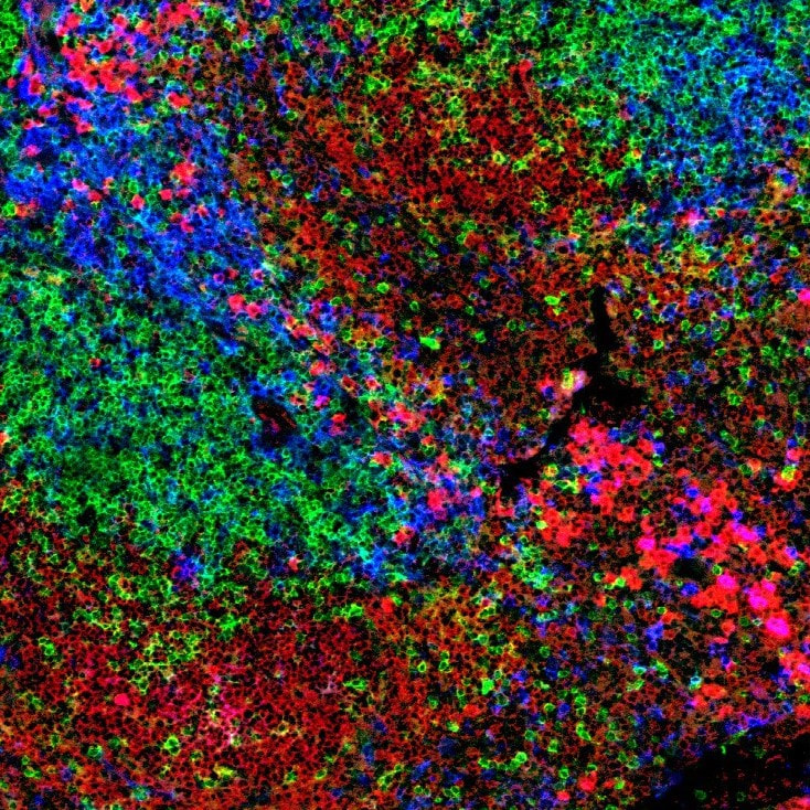
"Confocal image of the spleen of a mouse showing the B cells in red (stained for IgM), the helper T cells in green (stained for CD4) and the dendritic cells in blue (stained for CD11c)." Image by Peter Lane and Fiona McConnell has been cropped and is used under a CC BY 4.0 license.
|
REFERENCES AND RESOURCES
|
|
  |
